45 colon diagram with labels
Colon (Large Intestine): Anatomy, Function, Structure Sigmoid colon: The S-shaped connection between the last part of the colon and the rectum, located on the bottom left side of the abdomen is called the sigmoid colon. 2 Size and Length This organ is called the large intestine because of the diameter (width) of the intestine; it is much wider than the small intestine, but also much shorter. Understanding the Human Stomach Anatomy With Labeled Diagrams The former is located on the left, at the level of the tenth thoracic vertebra (T10). The pyloric orifice lies at the level of the first lumbar vertebra (L1). It is the opening from where the food travels towards the duodenum. Given below is a labeled diagram of the stomach to help you understand stomach anatomy. The stomach is divided into ...
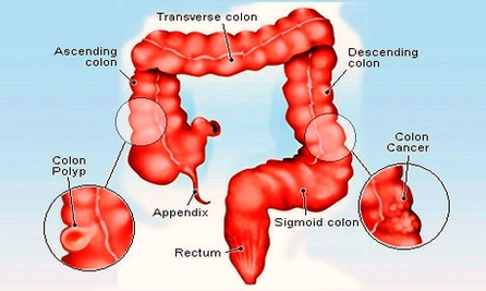
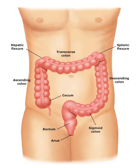
Anatomy of Colon and Rectum | SEER Training Anatomy of Colon and Rectum. The entire colon is about 5 feet (150 cm) long, and is divided into five major segments. The rectum is the last anatomic segment before the anus.. The ascending and descending colon are supported by peritoneal folds called mesentery.. The right colon consists of the cecum, ascending colon, hepatic flexure and the right half of the transverse colon.
Colon diagram with labels
Colon Anatomy - Human Body Diagrams - Medical Art Library Aug 28, 2017 · The large intestine begins at the cecum. The ileum (small intestine) ends where it connects to the cecum at the ileocecal junction. The colon is divided into four parts: the ascending, transverse, descending and sigmoid. The ascending and transverse colon meet at the right hepatic flexure (near the liver). The transverse and descending colon ... Labeled Diagram Of The Digestive System Stock Photos, Pictures ... Browse 116 labeled diagram of the digestive system stock photos and images available, or start a new search to explore more stock photos and images. ... Large Intestine, Colon Sections Labeled in Male Abdominal Anatomy labeled diagram of the digestive system stock pictures, royalty-free photos & images. Colon: Anatomy, histology, composition, function | Kenhub Oct 28, 2021 · The colon forms part of the large intestine and extends between the caecum and the rectum. It is about 1.5 meters in length and consists of four parts: ascending transverse descending sigmoid colon You can recognize it easily through several distinct morphological features like semilunar folds and pouches called haustra.
Colon diagram with labels. Labeled Diagram of the Human Kidney - Bodytomy The renal medulla comprises a set of 8-18 conical structures called renal pyramids that are surrounded by the cortex. Portions of the cortex between two adjacent pyramids are termed as renal columns. Spread in these pyramids and the cortex, are the functional units callednephrons. The actual filtration of blood occurs in the nephrons. The Digestive Tract with Labels | Media Asset | NIDDK Illustration of the digestive system with parts labeled: mouth, esophagus, stomach, large intestine (colon), small intestine, ileum, rectum, and anus. Caption The digestive tract. Diseases or Conditions Digestive Diseases File Size 992 KB | 2260 x 2492 File Type JPG How does the bowel work? Bowel information with diagrams - Macmillan ... Diagram of the position and sections of the small bowel The colon The colon is divided into 4 sections. Ascending colon The first part of the colon is joined to the small bowel and goes up the right side of the abdomen (tummy). Transverse colon The second section goes across the abdomen from your right to your left side. Descending colon The Colon - Ascending - Transverse - Descending - TeachMeAnatomy The colon (large intestine) is the distal part of the gastrointestinal tract, extending from the cecum to the anal canal. It receives digested food from the small intestine, from which it absorbs water and electrolytes to form faeces. Anatomically, the colon can be divided into four parts - ascending, transverse, descending and sigmoid.
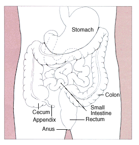
Large intestine with labels for the appendix, cecum ... The appendix, cecum, ascending colon, transverse colon, descending colon, sigmoid colon, rectum, and anus are labeled. Caption. The sigmoid colon is the last section of the colon. Diseases or Conditions. Digestive Diseases. File Size. 378 KB | 1192 x 1236. File Type. JPG. Related Keywords Colon (Large Intestine): Function, Anatomy & Definition Dec 8, 2021 — The large intestine includes the colon, rectum and anus. It's all one, long tube that continues from the small intestine and ends at the ... Colon Picture Labeling Flashcards - Quizlet Colon Picture Labeling STUDY Flashcards Learn Write Spell Test PLAY Match Gravity Created by XertziePLUS Terms in this set (11) rectum 10 external anal sphincter 12 teniae coli 14 cecum 8 vermiform appendix 9 transverse colon 2 ascending colon 5 ileum 6 ileocecal valve 7 sigmoid colon 13 descending colon 16 YOU MIGHT ALSO LIKE... Digestive System Labeled - Agaliprogram digestive system labeled. #1 (urine/pee pee) #2 (solid waste/poop) hydrochloric mechanical digestion chemical digestion saliva acid pepsin bile lipase stomach small intestine enzymes from liver and pancreas large intestine (transverse colon) descending coloncirculatory system there is an unlabeled diagram in the end of the article for readers to …
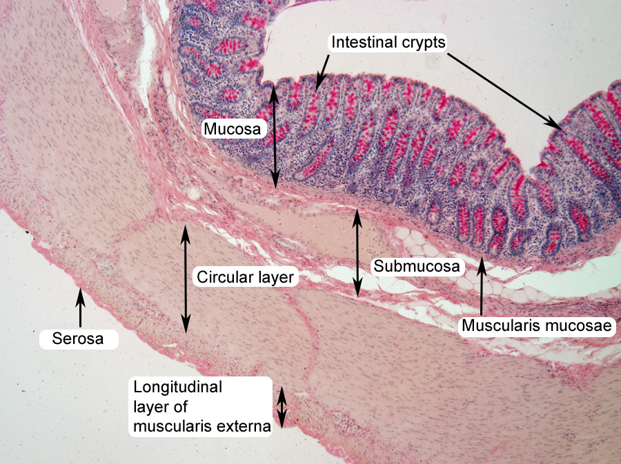
Intestine label Images, Stock Photos & Vectors | Shutterstock 1,953 intestine label stock photos, vectors, and illustrations are available royalty-free. See intestine label stock video clips ... Anatomy of the human ... Large Intestine Diagram - IvyRose Holistic Large labelled diagram of the anatomy of large intestine including the main ... The diagram of the large intestine (below) labels the features of the large ... PDF ANATOMIC DRAWINGS OF THE DIGESTIVE SYSTEM Esophageal sphincter Liver ... Colon (C18._).0 Cecum.1 Appendix.2 Ascending.3 Hepatic flex..4 Transverse.5 Splenic flex..6 Descending.7 Sigmoid.8 Overlapping.9 Colon, NOS Yes Yes Yes Yes Yes Yes Yes Yes Yes Yes Yes Yes Yes Yes Yes Yes Yes Yes Yes Yes Yes Yes Yes Yes Yes Yes Yes Yes Yes Yes Yes Yes Yes Yes Yes Yes Yes Yes Yes Yes Yes Yes Yes Yes Yes Yes Yes Yes Yes Yes Yes ... Colon Histology Slide with Labeled Diagram - AnatomyLearner Apr 06, 2022 · Colon Histology Slide with Labeled Diagram 04/06/2022 04/06/2022 by anatomylearner The colon histology slide possesses the typical four layers of a tubular organ – mucosa, submucosa, muscularis, and serosa. But, there are no permanent plica circularis and villi in the colon slide as found in the different segments of the small intestine.
Diagram of colon, rectum, and other parts of digestive system
Colonoscopy Measurements (cm) from Anal Verge | SEER Training Types of Surgery: Colon; Types of Surgery: Rectum; Radiation Therapy; Commonly Used Drugs; For hands-on exercises, please go to SEER*Educate. Resources. Archived Modules. Updates. Acknowledgements. Colonoscopy Measurements (cm) from Anal Verge. Return to Anatomy of Colon and Rectum. Follow SEER. Contact Information.
detail colon cancer | Anatomy System - Human Body Anatomy diagram and chart images
Parts of the colon | Understanding Your Colon or Rectal Surgery The colon (large intestine) is a long, muscular tube about four to five feet long. The colon removes water and nutrients from partially digested food. Then it turns the rest into stool (waste). The stool goes through the rectum and then leaves the body through the anus. Parts of the colon. Cecum: This is the beginning of the colon. It is ...

colon cancer | Anatomy System - Human Body Anatomy diagram and chart images
Illustration Picture of Anatomy - Colon - eMedicineHealth The colon is the largest part of the large intestine, extending from the cecum to the rectum. It is 5 feet long and its function is to reabsorb water from digested food and concentrate solid waste material, known as stool. The colon is made of several sections. The ascending colon travels up the right side of the abdomen.

57 best { Histology & A&P II } images on Pinterest
Picture of the Human Colon Anatomy & Common ... - WebMD The ileum (last part of the small intestine) connects to the cecum (first part of the colon) in the lower right abdomen. The rest of the colon is divided into four parts: • The ascending colon...
Digestive
PDF Digestive System Diagram Diagram . Large Intestine Mechanical Digestion Chemical Digestion Saliva Hydrochloric Acid Pepsin Trypsin Bile Lipase Stomach Small Intestine Enzymes from Liver and Pancreas Large Intestine (Transverse Colon) Descending Colon System Circulatory Kidneys #1 #2 Water and Vitamins Nutrients The Digestive System Esophagus Mouth On your Digestive ...

Gastrointestinal Tract - Colon Histology - Embryology
Colon Anatomy (with Small Intestine Label) - NCI Visuals Online 720x602. View. Download. Title: Colon Anatomy (with Small Intestine Label) Description: Drawing shows the cecum, ascending colon, transverse colon, descending colon, sigmoid colon, rectum, and anal canal. Also shown is the small intestine. The cecum connects the small intestine to the colon.
The Battle for Colonic Microflora | HIV and ID Observations
Intestines (Anatomy): Picture, Function, Location, Conditions The intestines include the small intestine, large intestine, and rectum. The small intestine (small bowel) is about 20 feet long and about an inch in diameter. Its job is to absorb most of the ...

The Polygon Monkey: Digestive System done in Modo and ZBrush
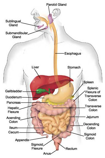
Abdomen and digestive system anatomy: diagrams labeled Full labeled anatomical diagrams - Anatomy of the abdomen and digestive system: these general diagrams show the digestive system, with the major human anatomical structures labeled (mouth, tongue, oral cavity, teeth, buccal glands, throat, pharynx, oesophagus, stomach, small intestine, large intestine, liver, gall bladder and pancreas).

Human Muscles Diagram / The Human Muscular System Anatomy Detailed Diagram 20 ...
Label Digestive System Diagram Printout - EnchantedLearning.com Read the definitions below, then label the digestive system anatomy diagram. anus - the opening at the end of the digestive system from which feces (waste) exits the body. appendix - a small sac located on the cecum. ascending colon - the part of the large intestine that run upwards; it is located after the cecum.

What Does the Colon do in the Digestive System
Colon Diagram Stock Illustrations - 3,263 Colon Diagram Stock ... Download 3,263 Colon Diagram Stock Illustrations, Vectors & Clipart for FREE or amazingly low rates! New users enjoy 60% OFF. 186,403,976 stock photos online. ... Labeled Diagram. Human colon. Colon - lymphatic drainage. Pathways of lymphatic drainage of the colon. Anatomy of the Colon, Rectum and Anus. Digestive system, large intestine ...
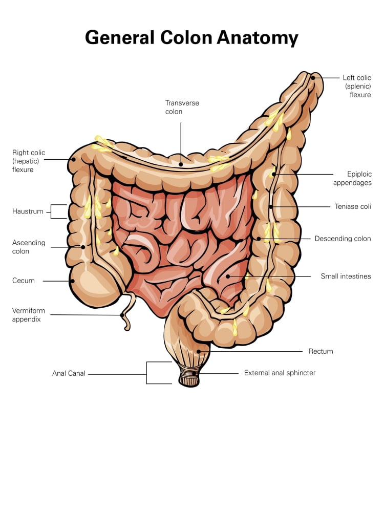
General colon anatomy, with labels. Poster Print by Alan Gesek/Stocktrek Images (24 x 32 ...
Large Intestine Anatomy, Parts, Diagram & Major Function - Study.com Draw a diagram of the large intestine and label all parts. Updated: 07/22/2021 Table of Contents. What is a Large Intestine? ... The three parts of the colon are the ascending colon, ...
Patient Resources | Gastroenterology and Hepatology
Sigmoid colon - Definition, Anatomy and Function | Kenhub Sigmoid colon - ventral view. The gastrointestinal system is divided into the foregut, midgut and hindgut.The foregut stretches from the oesophagus to the major duodenal papilla, the midgut from the major duodenal papilla to two thirds of the transverse colon, and the hindgut from this point to the pectinate line of the rectum.. Neurovasculature. The hindgut gets its blood supply from the ...

Human Digestive System Tract Stock Illustration - Download Image Now - iStock
Histology | Colon The 4 basic layers of the colon: This diagram illustrates the 4 basic layers of the colon. The inner pink layer is the mucosa, the yellow layer beneath the mucosa is called the submucosa, while the red layer is the muscular layer (muscularis) and the 4 th. layer is called the serosa or adventitia.. Courtesy Ashley Davidoff MD
Post a Comment for "45 colon diagram with labels"